
Berikut adalah gambar-gambar foto yang memenangi pertandingan Olympus BioScapes 2010. Sungguh unik sekali alam halus ciptaan Allah S.w.t. Walaupun teramat halus dari pandangan mata kasar, tetapi mempunyai corak-corak dan formasi-formasi yang amat canggih dan cantik. Hayatilah keindahan dan keunikan makhluk-makhluk ciptaan Allah ini yang telah dibesarkan menggunakan MIKROSKOP. Subhanallah .......
The Olympus BioScapes International Digital Imaging Competition honors the world’s most extraordinary microscope images of life science subjects.
10 - Weevil Head

This is the 10th-place picture, by British photographer Laurie Knight, shows the face of a weevil (possibly Curculio nucum or Curculio glandium). The image was captured using a lighting technique known as episcopic illumination.
9 - Wildflower seeds

China’s Yanping Wang won ninth place in the 2010 Olympus BioScapes competition for this image of wildflower seeds, captured using brightfield reflected light.
8 - Beetle Leg

German researcher Jan Michels’ eighth-place image shows a lateral view of the adhesive pad on the leg of a beetle (Clytus sp.). The view was captured using autofluorescence.
7 - Damselfly’s Eye

Germany’s Igor Siwanowicz won seventh place in the Olympus BioScapes competition for this view of the eye of a damselfly. This projection of a series of confocal microscope images shows the regular, crystal-style architecture of the eye of Enallagma cyathigerum. The area covered in the photo measures about 0.6 by 0.8 millimeters, or 0.02 by 0.03 inches.
6 - Spirogyra

Polish biotech researcher Jerzy Gubernator took this extreme close-up of Spirogyra algae using brightfield and polarized light. The photomicrograph won the sixth-place prize in the 2010 Olympus BioScapes competition.
5 - Primordium
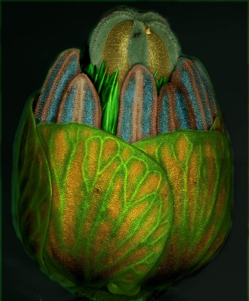
This picture by Iranian horticulturist M. Reza Dadpour shows the primordium (bud) of the weedy flower Tribulus sp. during its final stages of development. More than 100 images, focused at different depths, were composed to produce this view. The image won fifth prize in the 2010 Olympus BioScapes competition.
4 - Diatoms

Fanlike diatoms, representing the species Licmophora juegensii, have latched onto red algae in this fourth-place picture by Germany’s Wolfgang Bettighofer. Licmophora cells are able to move and, supported through light sensors, locate a place with suitable light exposure. Then they produce a sticky stalk that keeps them attached to their home base. The sample was collected from brackish water in the Baltic Sea.
3 - Coral

The third-place image in the Olympus BioScapes competition shows a solitary coral (Fungia sp.). The tentacle tips, called acrospheres, are visibly enhanced. This picture was taken by James Nicholson, a coral researcher in South Carolina.
2 - Rat Brain

California’s Thomas Deerinck won second place in the Olympus BioScapes competition for this image of a structure known as the hippocampus, within the brain of a rat. The tissue was stained to reveal the distribution of glial cells (blue), neurofilaments (green) and cell nuclei (yellow).
1 - Daddy Longlegs

Germany’s Igor Siwanowicz won first place in the Olympus BioScapes competition for this image, showing the eyes of a spiderlike bug known as a Daddy Longlegs or Harvestman. The picture has been color-coded to reflect depth, and shows the lenses (two large ovals), the retinas and the optic nerves
Tiada ulasan:
Catat Ulasan